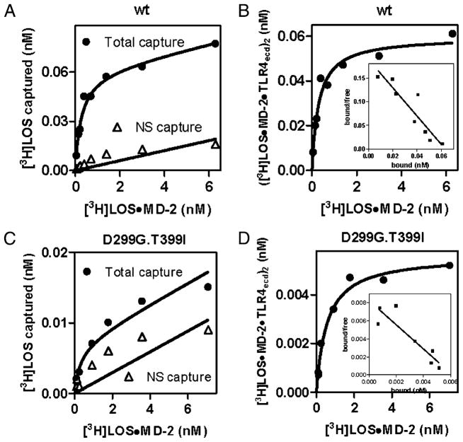
FIGURE 2.
Comparison of dose-dependent binding of [3H]LOS·MD-2 to soluble TLR4ecd (wt [A, B] versus D299G.T399I variant [C, D]). Increasing concentrations of [3H]LOS·MD-2 were incubated for 30 min at 37°C in PBS/0.1% HSA supplemented with conditioned medium from HEK293T cells expressing FlagTLR4ecd (wt or D299G.T399I) or from mock-transfected cells. Cocapture of [3H]LOS with ANTI-FLAG affinity gel was measured as described in Materials and Methods and data were analyzed as described in the legend to Fig. 1. Data are from one experiment, representative of three closely similar experiments.

