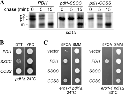
FIGURE 8.
Pdi1p a domain mutant shows diminished oxidative folding capacity. A, processing of CPY in pdi1Δ cells containing PDI1 plasmids. Cells were pulse labeled for 7 min and chased at 30 °C, and CPY was immunoprecipitated and resolved by SDS-PAGE. ER (p1), Golgi (p2), and vacuolar (m) forms of CPY are indicated. B, yeast strains from panel A were grown for 12 h in SMM without leucine and spotted onto YPD plates with or without 10 mm DTT. Strains were grown for 2 days at 24 °C. C, the pdi1Δ strain CKY1058 covered by an URA3-marked PDI1 plasmid was transformed with LEU2-marked plasmids encoding Pdi1p (pCS463), Pdi1p(SSCC) (pCS465), Pdi1p(CCSS) (pCS464), or empty vector. These strains were spotted onto SMM plates or SMM plates containing 5-fluoroorotic acid (to select for yeast able to grow in the absence of the URA3 plasmid). Plates were incubated at 24 °C, 30 °C, or 37 °C for 3 days. All four strains were inviable at 37 °C (not shown), consistent with the previously characterized lethality of the ero1-1 mutant at 37 °C (1).

