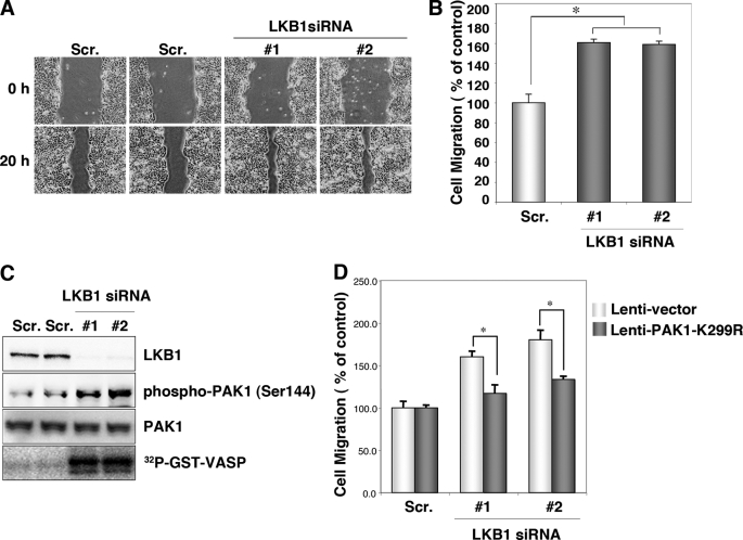
FIGURE 1.
Knockdown of endogenous LKB1 increases cell migration and PAK1 activity in HCT116 cells. A, knockdown of LKB1 increases cell migration. HCT116 cells were transfected with two different specific siRNAs against human LKB1. Control cells were transfected with a scrambled (Scr.) siRNA pool. After 24 h of plating, scratches were made with 10-μl pipette tips. Photographs were taken at 0 and 20 h after the wound was made. B, cell migration was normalized so that 100% represented migration distance of control cells. Error bars indicate S.D. The asterisk indicates significant increases compared with control cells (p < 0.001). C, cellular levels of phospho-PAK1 (Ser144), total PAK1 and in vitro PAK1 activity in LKB1 knockdown HCT116 cells. Lysates were prepared from HCT116 cells transfected with the indicated siRNAs. The levels of phospho-PAK1 (Ser144) and PAK1 were determined by Western blotting with the indicated antibodies. Lysates were prepared from HCT116 cells transfected with the indicated siRNAs and immunoprecipitated using an anti-PAK1 antibody. The precipitates were used for in vitro kinase assays using GST-VASP-(158–277) as a substrate. The phosphorylation of GST-VASP was visualized using BAS-5000 Bio-imaging Analyzer. D, induction of PAK1-K299R suppressed the increase in migration in LKB1 knockdown cells. The cells transfected with the indicated siRNAs were then infected with lentivirus lenti-PAK1-K299R or lenti-vector. At 24 h post-infection, cultures were scratches were made with 10-μl pipette tips. Photographs were taken at 0 and 20 h thereafter. Cell migration was normalized so that 100% represented the migration distance of control cells. Error bars indicate S.D. The asterisk represents significant increases compared with control cells (p < 0.001).

