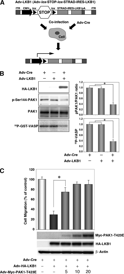
FIGURE 2.
Expression of LKB1 in the Lkb1-null MEF cell line (MEF3-2) inhibits cell migration and PAK1 activation. A, schematic structure of the adenoviral construct, adapted from Ref. 17. ITR, inverted terminal repeat; CMV, human cytomegalovirus immediate early promoter; lox, loxP site sequence; STOP; transcriptional termination cassette; IRES, internal ribosome entry site; pA, polyadenylation signal. Co-infection with Adv-Cre removed the STOP cassette from Adv-LKB1. B, cellular levels of PAK1 autophosphorylation, total PAK1, and in vitro PAK1 activity in LKB1-expressing MEF3-2 cells. MEF 3-2 cells were co-infected with recombinant adenoviruses Adv-Cre and Adv-LKB1. Control cells were infected with Adv-Cre alone. The levels of phospho-PAK1 and PAK1 were determined by Western blotting with anti-phospho-PAK1 (Ser144)/PAK2 (Ser141) and anti-PAK1 antibodies, respectively. Lysates were prepared from MEF3-2 cells infected with the indicated recombinant adenoviruses and immunoprecipitated using an anti-PAK1 antibody. The precipitates were used for in vitro kinase assays with GST-VASP-(158–277) as a substrate. The phosphorylation of GST-VASP was visualized and quantified using BAS-5000 Bio-imaging Analyzer. Error bars indicate S.D. The asterisk represents significant decreases compared with control cells (p < 0.001). C, the constitutively active mutant of PAK1 rescues LKB1-induced suppression of cell migration in MEF3-2 cells. MEF 3-2 cells were co-infected with recombinant adenoviruses Adv-Cre, Adv-LKB1, and Adv-PAK1-T423E at a multiplicity of infection of 5, 10, or 20. Control cells were infected with Adv-Cre alone. Cell migration assays were performed as Fig. 1. Error bars indicate S.D. The asterisk represents significant changes in Adv-Cre/Adv-LKB1/Adv-PAK1-T423E (5) infected cells compared with Adv-Cre/Adv-LKB1-infected cells (p < 0.001). Similar results were obtained in three independent experiments.

