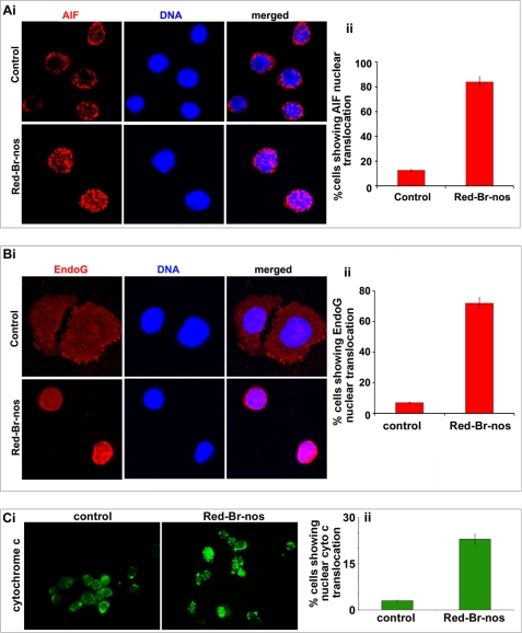
FIGURE 4.
Red-Br-nos treatment induces caspase-independent cell death in PC-3 cells. Micrographs show immunocytochemical staining of 48 h Red-Br-nos-treated PC-3 cells for AIF (panel A, i), Endo-G (panel B, i), and cytochrome c (panel C, i). Bar graph representation of the quantitation of percent cells showing nuclear AIF translocation (panel A, ii), nuclear Endo-G translocation (panel B, ii) and nuclear cytochrome c translocation (panel C, ii) in control and drug-treated PC-3 cells from 6 to 8 random image fields totaling 200 cells and reported as mean ± S.D. (p < 0.05, compared with controls).

