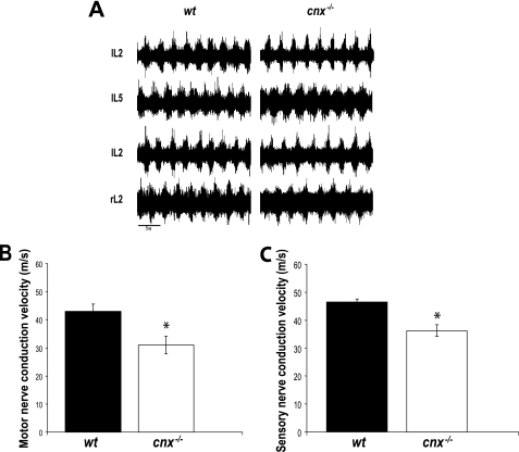
FIGURE 3.
Functional analysis of calnexin in neuronal tissue. A, the fictive locomotor pattern is undisturbed in the cnx−/− mouse. Electroneurograms were recorded from the second and fifth lumbar ventral root on the left side (i.e. IL2, IL5) and the second lumbar ventral root on the left and right side (i.e. IL2, rL2) of the spinal cord in the wild-type (wt, left traces) and cnx−/− (right traces) mice. Fictive locomotion was evoked with 5 μm serotonin (5-hydroxytryptamine) and 10 μm N-methyl-d-aspartic acid. The spinal cords used were obtained from newborn, 1-day-old, and 2-day-old mice (n = 4). Note the appropriate alternation between bursts in both cases. B, motor nerve conductive analysis of wild-type (wt) and calnexin-deficient (cnx−/−) mice. Motor nerve conduction velocity was significantly slower in the absence of calnexin. C, sensory nerve conductive analysis of wild-type (wt) and calnexin-deficient (cnx−/−) mice. In the absence of calnexin, there was reduced sensory nerve conduction velocity. Six sex-matched sets of wild-type and calnexin-deficient mice of 4–5 months of age were examined for motor and sensory nerve conduction. The asterisks indicate significant differences (p = 0.01).

