
Figure 1. ERβ1 and EMT of PCa.
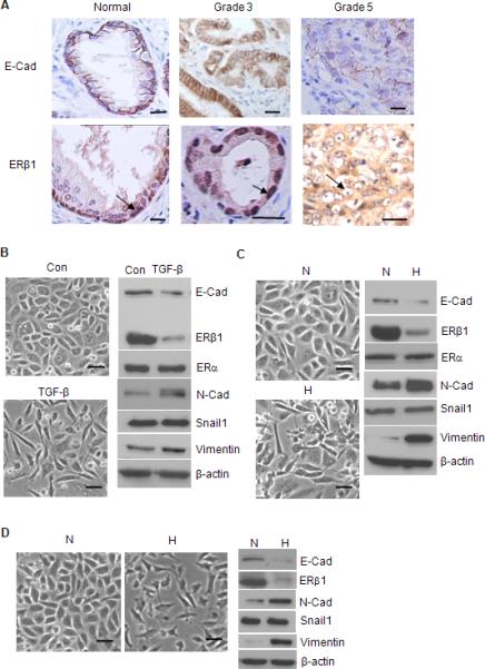
(A) Specimens of normal glandular epithelium, Gleason grade 3 and 5 PCa were stained for E-cadherin and ERβ1 and photographed. ERβ1 is localized in the nuclei of basal cells in the normal prostate and nuclei of grade 3 tumor cells (arrow). In contrast, nuclear ERβ1 staining is absent in grade 5 PCa (arrow). The data are representative of 3 separate specimens for each classification. Scale bars = 20 μm. (B) PC3 cells were treated with PBS (con) or TGF-β for 3 days, photographed and extracts were analyzed for the expression of EMT markers and ERβ1 by immunoblotting. PC3 (C) or LNCaP (D) cells were maintained in either normoxia (N) or hypoxia (H) (0.5% O2) for 24 hrs, photographed and extracts from these cells were immunoblotted as described above. Scale bars = 50 μm. See also Figure S1.