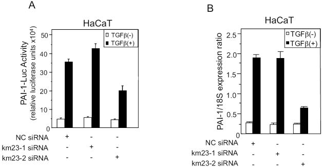
Fig. 3. TGFβ-mediated induction of PAI-Luc luciferase activity and endogenous PAI-1 gene expression in HaCaT cells specifically requires km23-2, but not km23-1.
A: HaCaT cells were transfected with NC siRNA, km23-1 siRNA, or km23-2 siRNA. 24h after transfection, the medium was replaced with SF medium for 1 h, followed by incubation of cells in the absence (open bar) and presence (black bar) of TGFβ (5ng/ml) for an additional 18 h. Luciferase activity was measured using the Dual Luciferase Reporter Assay System (Promega). The results are representative of at least two experiments, each performed in triplicate. B: HaCaT cells were transfected and treated as described for Fig. 3A. Real-time RT-PCR analysis of human PAI-1 mRNA expression from HaCaT cells was performed. Bars represent mean ± SD of PAI-1 mRNA levels normalized to control 18S rRNA levels. Results are representative of two experiments.

