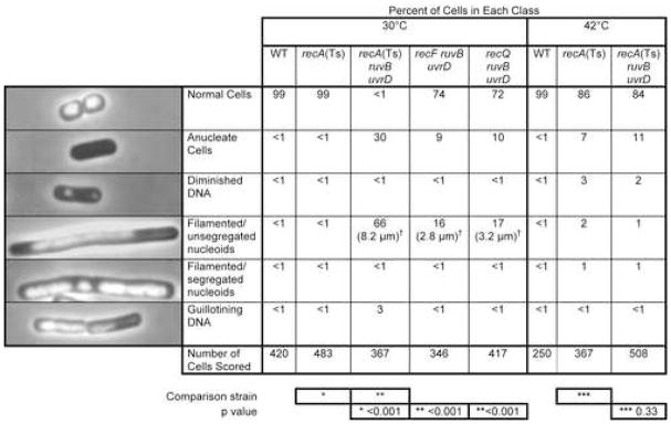
Figure 3.
Chromosome-Segregation Failure Accompanies Death of Δruv ΔuvrD Cells
Cultures were grown from common 42°C-grown saturated cultures, split, diluted and grown to log-phase at 30°C or 42°, harvested, prepared for DAPI DNA-fluorescence microscopy, and scored blind. p values calculated from Chi-squared analyses indicate significant differences in the distributions between the indicated strains (* versus *, ** versus **, etc.) (SigmaStat 3.1 from SPSS, Inc). Table S3 for comparisons of numbers of normal cells in all relevant single- and double-mutant combinations.
†Median cell lengths. ~2.7-fold reductions by ΔrecF and ΔrecQ compared with the recA(Ts)ΔruvB ΔuvrD cells. The median length of 100 wild-type (normal) cells was 1.5 μm.

