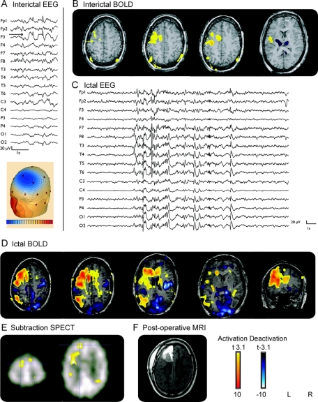
Figure 1 Patient 2
(A) Interictal EEG (average montage): focus F3. (B) Interictal functional MRI (fMRI): positive blood oxygen level dependent (BOLD) response: left superior and middle frontal gyri; negative BOLD response: thalamus. (C) Ictal EEG: polyspikes without lateralization. (D) Ictal fMRI: positive BOLD responses same as interictal but more extended including left cingulate gyrus and left parietal; negative BOLD response: thalamus, occipital, frontobasal, parietal. (E) Subtraction of interictal and ictal SPECT showing a left frontal focus. (F) Postoperative MRI after frontoparamedian resection.

