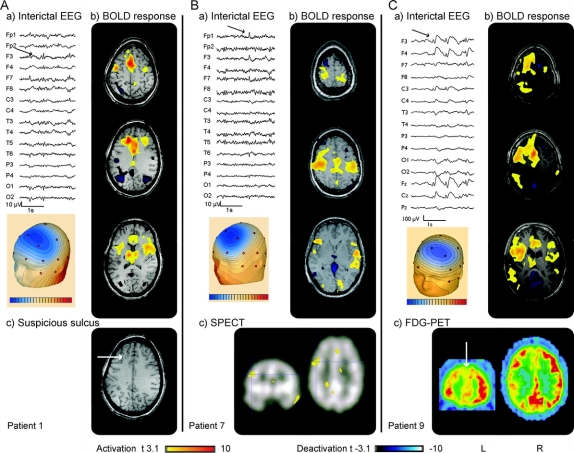
Figure 3 Patient 1
(A) EEG (average montage) focus F3, F7. (B) Functional MRI (fMRI): maximum of positive blood oxygen level dependent (BOLD) response left middle frontal gyrus; negative BOLD response bilateral in the parietal cortex. (C) MRI: suspicious deep left middle frontal sulcus. Patient 7: (A) EEG (average montage) focus F3, F7. (B) fMRI: positive BOLD response: bilateral motor cortex, unrelated to spike topography; negative BOLD response: precuneus. (C) Subtraction of interictal and ictal SPECT showing a focus frontolateral left. Patient 9: (A) EEG (average montage) focus F3, Fz, with frequent generalization. (B) fMRI: maximum of positive BOLD response left frontomesial, additionally frontolateral, bilateral parietal left more than right; negative BOLD response: precuneus, occipital cortex, and caudate nuclei. (C) FDG-PET: hypometabolism left frontal cortex, extended to parietal and temporal.

