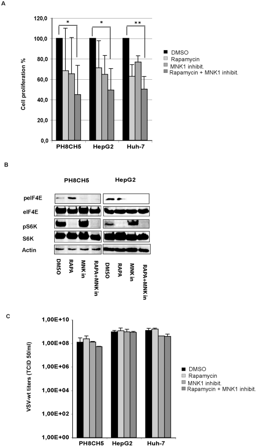
Figure 11. Effects of concomitant inhibition of mTOR and MNK on VSV proliferation.
Cells were mock-treated (DMSO) or treated with rapamycin at 50 nm, MNK1 inhibitor at 20 µM, alone or together as indicated. A) Cell proliferation assays were performed using the MTT assay. Representative results of at least two independent experiments are shown. B) Western blot analysis of lysates obtained by PH5CH8 and HepG2 cell lines mock-treated (DMSO) or treated with rapamycin (RAPA), MNK inhibitor (MNK in) alone or in combination (RAPA+MNK in). The levels of S6K and eIF4E phosphorylated forms were monitored after inhibitor treatment. C) Cells were infected with VSV-wt at an MOI of 0.1 for 24 hours. Viral titers represent the mean ± standard deviation of three experiments.

