
FIGURE 1.
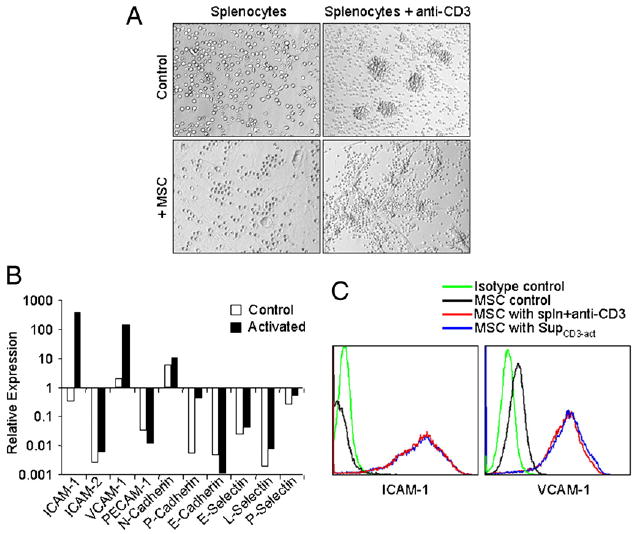
Expression of ICAM-1 and VCAM-1 in MSCs was greatly induced by T cell products. A, Fresh C57BL/6 splenocytes were stimulated with or without anti-CD3 (1 μg/ml) and cultured in the presence or absence of MSCs derived from C57BL/6 mice at a 20:1 ratio (splenocytes/MSCs). The cells were examined microscopically after 48 h (original magnification ×200). Representative of 10 independent experiments. B, Expression of adhesion molecules at mRNA levels in MSCs cocultured with activated splenocytes. MSCs were incubated for 48 h in the presence of fresh splenocytes activated by anti-CD3 (1 μg/ml). MSCs not exposed to activated splenocytes were used as controls. After removing the nonadhesive lymphocytes, the MSCs were purified by a CD45-microbeads kit using a negative selection-based MACS sorting. The gene expression of the adhesion molecules in purified MSCs (>95% pure based on flow cytometry test) and the control MSCs were analyzed by real-time PCR and compared with β-actin mRNA, defined as 1000 arbitrary unit. Representative of three independent experiments. C, Expression of ICAM-1 and VCAM-1 assayed by flow cytometry. MSCs were cultured with or without fresh splenocytes as in B or supplemented with SupCD3-act (50% of total volume) for 24 h. The expression of ICAM-1 and VCAM-1 in MSCs was detected by flow cytometry using electronic gating to exclude lymphocytes. Untreated MSCs served as a control. Data shown are mean ± SD of a representative of five experiments.