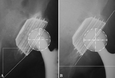
Fig. 3A–B.
The radiographic view of a cup placed at an extremely superolateral position and radiographic schedules of Roman software program for measurement of mean linear PE wear rate are shown. (A) An AP radiographic view of a ceramic-on-PE cup taken 1 month postoperatively shows the cup placed at a superior and lateral position out of the Ranawat triangle with a partial cranial gap and gap between the front end of the implant and the acetabular floor as a result of the surgical technique. (B) An AP radiographic view of the cup taken 9 years postoperatively shows a stable metal cup shell, periprosthetic osseointegration, and no measurable PE wear.

