
Fig. 3.
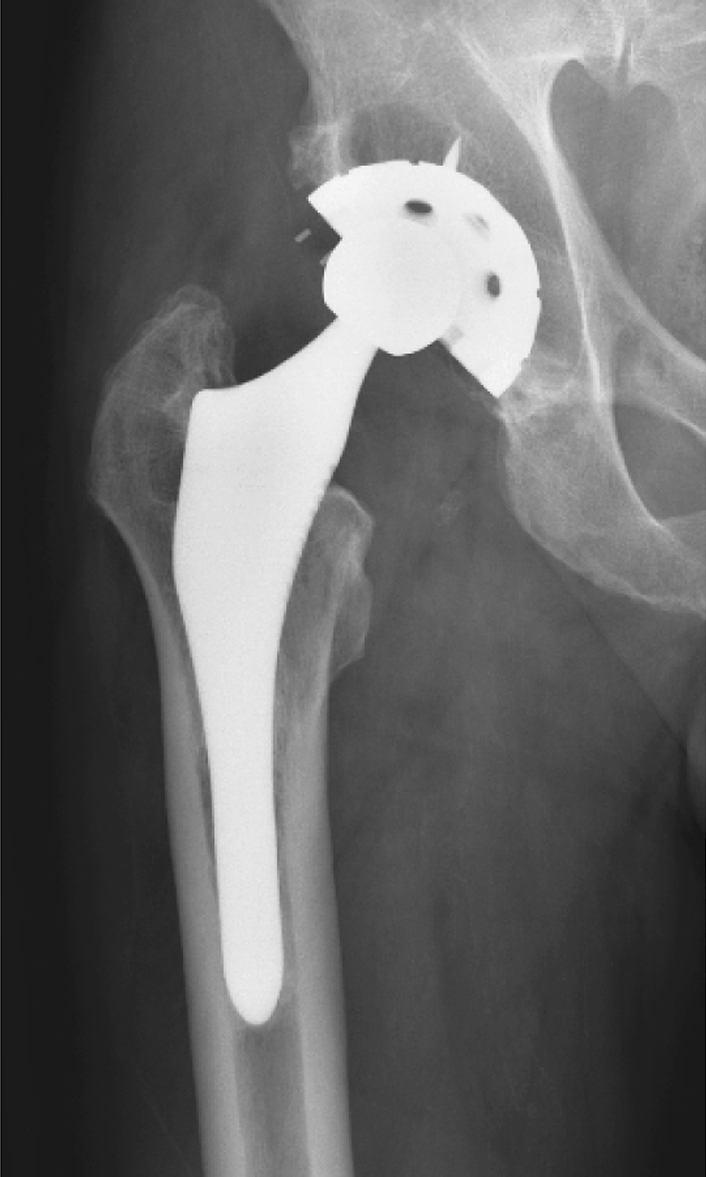
An AP radiograph of a surviving ABG I prosthesis shows evidence of osteolysis in all three DeLee and Charnley acetabular zones. The osteolysis in Zones 2 and 3 is of low volume and this implant was considered well fixed. This patient had an OHS of 14 of 60 at 13 years postoperation.