
Fig. 1.
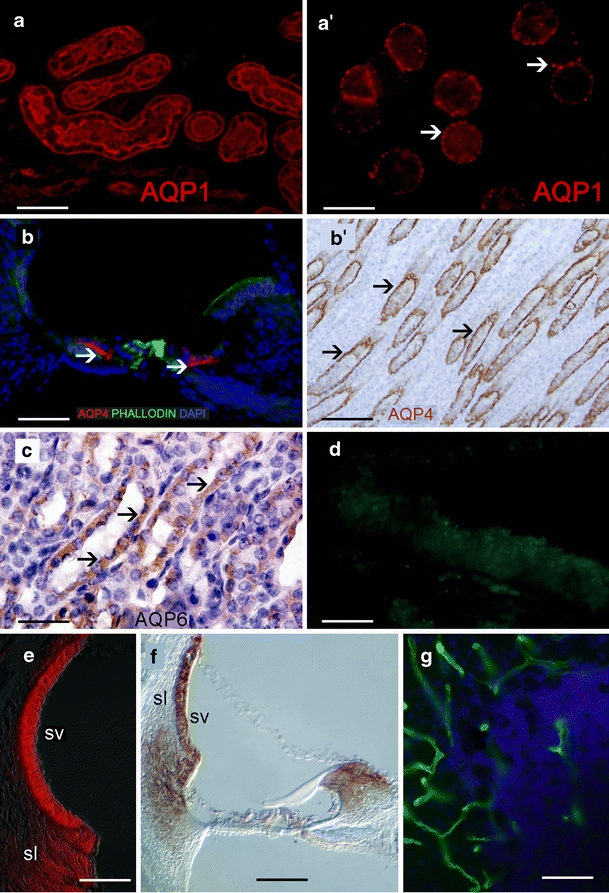
Immunohistochemical controls. a AQP1 immunoreactivity (red) in the human kidney. a’ AQP1 immunoreactivity (red) in human red blood cells. AQP1 localizes on the membrane (arrowheads). b AQP4 immunoreactivity (red) in a cross section of the rat organ of Corti. Inner sulcus and outer sulcus cells are AQP4-immunoreactive (arrowheads). Phalloidin Oregon green staining was used to identify actin in the apical portion of supporting cells (green). b’ AQP4 immunoreactivity in the rat kidney (amber) counterstained with hematoxylin (blue nuclei). c AQP6 immunoreactivity in the rat kidney (amber, arrows) counterstained with hematoxylin (purple nuclei). d Cross section of a human macula utriculi from an autopsy; AQP1 was absorbed with the corresponding peptide. No specific immunoreaction was detected. e Na+K+ATPase (red) in the rat stria vascularis (SV) and spiral ligament (sl). f NKCC1 in the rat SV, sl, and spiral limbus (right; dark amber). g α-Syntrophin in blood vessels of the mice cerebellum (green). Bars 50 µm (a, b, e, f), 10 µm (a’), 120 µm (b’), 40 µm (c), 45 µm (d), 25 µm (g)