
Fig. 2.
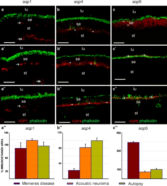
AQP 1 (aqp1), AQP4 (aqp4), and AQP6 (aqp6) immunoreactivity in the utricular macula of Meniere’s, acoustic, and autopsy patients. AQP1 immunoreactivity (red) in Meniere’s disease (a), acoustic neuroma (a’), and autopsy specimens (a’’) is localized to the fibrocytes beneath the overlying sensory epithelium (se, arrow) and to the fibrocytes in the underlying stroma (st, double arrows) in the utricular maculae (lu luminal region). AQP1 immunoreactivity was not present in the sensory epithelium itself. A quantitative comparison of AQP1 immunoreactivity between the three types of specimens (a’’’) showed no statistically significant differences. AQP4 immunoreactivity (red) in Meniere’s disease (b), acoustic neuroma (b’), and autopsy specimens (b’’) was localized to the basal pole of supporting cells. The AQP4 immunoreactivity in the utricular macula from subjects with Meniere’s disease (b) was significantly diminished in the sensory epithelium (arrows) when compared with that in acoustic neuroma (b’, arrows) and in autopsy material (b’’, arrows). Quantitative immunoreactivity analysis (b’’’) shows a statistically significant decrease in AQP4 immunoreactivity in Meniere’s specimens when compared with acoustic neuroma and autopsy specimens. AQP6 immunoreactivity in Meniere’s disease (c), acoustic neuroma (c’), and autopsy specimens (c’’). In the utricular macula from subjects with Meniere’s disease (c), AQP6 immunoreactivity (red) was diffusely distributed throughout the supporting cell in the sensory epithelium (arrows). In utricular maculae from patients with acoustic neuroma (c’) and normative subjects (c’’), AQP6 immunoreactivity was polarized to the sub-apical portion of the supporting cells. An increased intensity and more diffuse expression of AQP6 (red) was noted in Meniere’s disease (c) compared with acoustic neuroma (c’) and normative (c’’) material. Quantitative immunoreactivity analysis (c’’’) shows a statistically significant increase in AQP6 immunoreactivity in Meniere’s specimens when compared with acoustic neuroma and autopsy specimens. Phalloidin Oregon green staining (green) was used to identify actin at the apical portion of the sensory epithelium. No co-localization was observed between AQP6 (red) and phalloidin (green) corroborating a sub-apical distribution in the supporting cell of utricular maculae derived from acoustic neuroma (b) and normative (c) samples. Bars 50 µm (a–a’’, b–b’’, c–c’’)