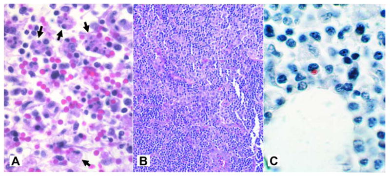
Figure 12.

A) Hemophagocytosis (arrows) in lymph nodes from a patient with HME (H&E; original magnification 240×). B) High cellularity in lymph node from patient with HME (magnificationX 64). C) Immunohistochemical staining of lymph nodes from patient with HME shows typical low bacterial burden (immunoperoxidase with hematoxylin counterstain; original magnification 240×). (From Dierberg KL, Dumler JS. Lymph node hemophagocytosis in rickettsial diseases: a pathogenetic role for CD8 T lymphocytes in human monocytic ehrlichiosis (HME)? In: BMC Infect Dis. 2006; 6:121. This is an Open Access article distributed under the terms of the Creative Commons Attribution License (http://creativecommons.org/licenses/by/2.0), which permits unrestricted use, distribution, and reproduction in any medium, provided the original work is properly cited.)
