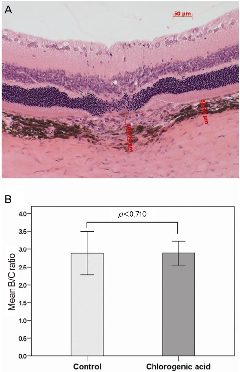
Fig. 2.
(A) In order to evaluate changes in the choroidal neovascularization lesion, the ratios of B (the thickness from the bottom of the pigmented choroidal layer to the top of the neovascular membrane) to C (the thickness of the intact-pigmented choroid adjacent to the lesion) were compared. (B) There was no significant difference in the B/C ratio between the chlorogenic acid-treated and control groups.

