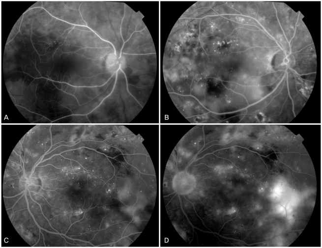
Fig. 2.
Fluorescein angiography (FAG) of both eyes revealed numerous fluorescent blotches in the subretinal space. FAG demonstrated hypofluorescence in the early phase and hyperfluorescence in the late phase resulting from leakage of fluorescein dye into the subretinal space, in addition to multiple microaneurysms resulting from diabetic retinopathy. (A) Early phase (right eye). (B) Late phase (right eye). (C) Early phase (left eye). (D) Late phase (left eye).

