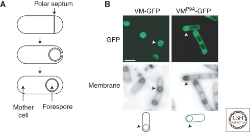
Figure 3.
Asymmetric localization of SpoVM. (A) Stages of sporulation. (Top) Division creates a mother cell and a smaller forespore. (Middle) The mother cell engulfs the forespore. (Bottom) The forespore is pinched off as a protoplast. (B) SpoVM-GFP localizes to the surface of the forespore, whereas SpoVMP9A-GFP localizes to all membranes. Arrowheads identify the cell depicted in the illustrations. Illustration provided by Kumaran Ramamurthi.

