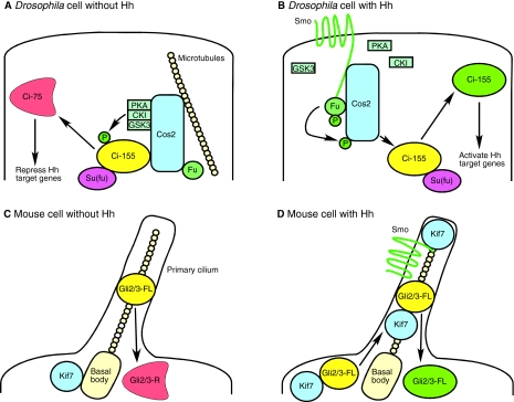
Fig. 4.
Hedgehog signaling complex function in Drosophila and mouse. Modes of action of the Hh signaling complex. Matching colors denote homologs. (A) In Drosophila, Cos2 associates with microtubules and scaffolds PKA, CKI and GSK3. This leads to efficient phosphorylation of Ci-155 and limited proteolysis to generate Ci-75. (B) Cos2 binds to the C-tail of Smo when Smo is in its active conformation, leading to the dissociation of PKA, CKI and GSK3. Fu is phosphorylated and in turn phosphorylates Cos2, which might promote the dissociation of Ci-155, leading to activation of Hh target genes. Removal of components of the Hh signaling complex has variable effects on Ci-155 and Ci-75 levels, and the relationship of these protein levels to the activation or repression of Hh target genes is not completely understood. (C) In the mouse, Gli2 and Gli3 are present in small amounts on the primary cilium in the absence of Hh ligand, and the cilium is required for generation of Gli repressors. Kif7 is located at the base of the cilium. (D) Kif7 binds Gli proteins and regulates their translocation to the primary cilium after stimulation of a cell with Hh ligand. Cilia promote activation of Gli2 and Gli3 through unknown mechanisms. Kif7 might also bind Smo, and Smo is required for Kif7 to move up the cilia. The precise makeup of the vertebrate complex in the absence and presence of Hh remains to be determined, although genetic evidence demonstrates that Fu is not an essential part of it. Ci, Cubitus interruptus; CKI, casein kinase I; Cos2, Costal2; FL, full-length; Fu, Fused; GSK3, glycogen synthase kinase 3; Hh, Hedgehog; Kif7, kinesin family member 7; P, phosphate group; PKA, protein kinase A; R, repressor; Smo, Smoothened; Su(fu), Suppressor of fused.

