Fig. 2.
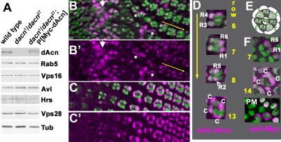
dAcn is expressed in the nuclei of eye disc cells. (A) In western blots of Drosophila third instar larval lysates, dAcn levels were reduced in dacn1/dacn27, but the levels of the other indicated proteins were similar to those in wild type. (B-D) Eye discs expressing nuclear GFP (green) were stained for dAcn (magenta). Posterior is to the right. dAcn accumulates in nuclei at the morphogenetic furrow and in a dynamic pattern in photoreceptor and cone cells. (B,C) Micrographs show cells either close to the furrow (B, white arrow) or cells more apical and more posterior (C). Asterisks mark examples of cells with cytoplasmic staining. The three ommatidia next to the yellow arrow are magnified in D, as labeled by the specific rows of their location. Photoreceptor or cone cells (c) with strongest staining in each ommatidium are indicated. (E) Schematic illustrating the relative positions of cells in ommatidia. (F) Eye disc expressing Myc-tagged dAcn was stained with anti-Myc antibodies to reveal dAcn expression in ommatidia of the indicated row or the peripodial membrane (PM). Genotypes are: (B-D) nGFP33 nGFP38 FRT40A/CyO; (F) dacn27 nGFP33 nGFP38 FRT40A/dacn1; P[genomic Myc-dacn].

