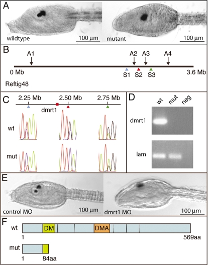
Fig. 1.
Anterior neural mutant maps to dmrt1 gene. (A) Trunk region of wild-type and homozygous ENU34 mutant at larval stage. (B) Diagram of Reftig 48 showing linked AFLP (A1-A4) and SNP (S1-S3) markers. (C) Linkage analysis with SNP markers S1, S2 and S3. Representative traces are shown. (D) RT-PCR for dmrt1 and laminin α3/4/5 (lam; used as a loading control) in wild-type and homozygous ENU34 mutants. (E) A translation-blocking MO to dmrt1 phenocopies the ENU34 mutant (compare right embryo in E with right embryo in A). (F) Diagram of the predicted wild-type and ENU34 mutant C. savignyi Dmrt1 protein.

