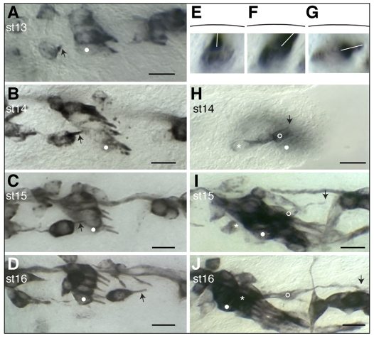
Fig. 3.
Morphogenesis and migration of the v'ch1 neuron and cap cell in wild-type Drosophila embryos. Single hemisegments of embryos were fixed at the developmental stage indicated. (A-G) mAb22C10-stained embryos showing v'ch1 dendrite morphology (arrow). (H-J) P0163-GAL4; UAS-tau-lacZ embryos stained with anti-β-galactosidase, showing morphology of the v'ch1 neuron (asterisk) and cap cell (open circle). White dots in A-D and H-J show the position of the lch5-5 neuron. The arrow in H-J shows the dorsal edge/tip of the v'ch1 cap cell. (E-G) Cross-sectional views of v'ch1 reconstructed from a z-series of images. The black line shows the position of the epidermis. The white line shows the dendrite direction. Scale bars: 10 μm.

