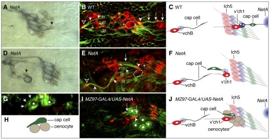
Fig. 7.
Aberrant morphogenesis of v'ch1 cap cell following loss of function or misexpression of Netrin-A. Images of stage-16 Drosophila embryos with the genotypes indicated. (A,D) The scolopale cell nucleus (arrow) is revealed by anti-Prospero immunohistochemistry (brown), and sensory neurons are labelled with mAb 22C10 (blue). The scolopale cell associates closely with the dendrite, whether it is in a normal (A) or aberrant (D) position. (B,E) Cap cells are stained with anti-β-tubulin (green) and neurons with anti-HRP (red). The v'ch1 neuron in the NetA embryo in E (outlined, not included in projection for sake of clarity) has stalled ventral to the lch5 cluster (asterisk); compare with its normal position in the wild-type embryo (B). The cap cell (large arrows) retains its association with the v'ch1 dendrite (arrowhead) in the mutant, but it is oriented anteroventrally and attaches to the epidermis near the cap cell of vchB (small arrows), close to the muscle 21 and 22 attachment sites. The vchB dendrite in B and E is indicated by an open arrowhead. (G,I) NetA was ectopically expressed in oenocytes (white dots) using the MZ97-GAL4 driver line. (G) A transverse view reveals that the cell body of the v'ch1 cap cell (asterisk) has remained in the epidermal layer, directly superficial to the oenocytes and has extended an aberrant ventral process (arrows) along the surface of an oenocyte. (I) The v'ch1 cap cell has projected an aberrant ventral process (arrows) but the nucleus has not translocated into this process. (C,F,H,J) Schematics of B,E,G,I are shown in C,F,H,J, respectively. The v'ch1 neuron cell body is outlined. The site of endogenous NetA expression is shown in blue in C and J. Scale bars: 10 μm.

