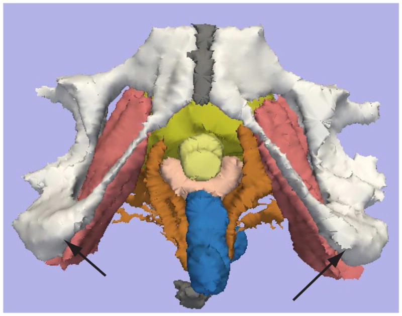
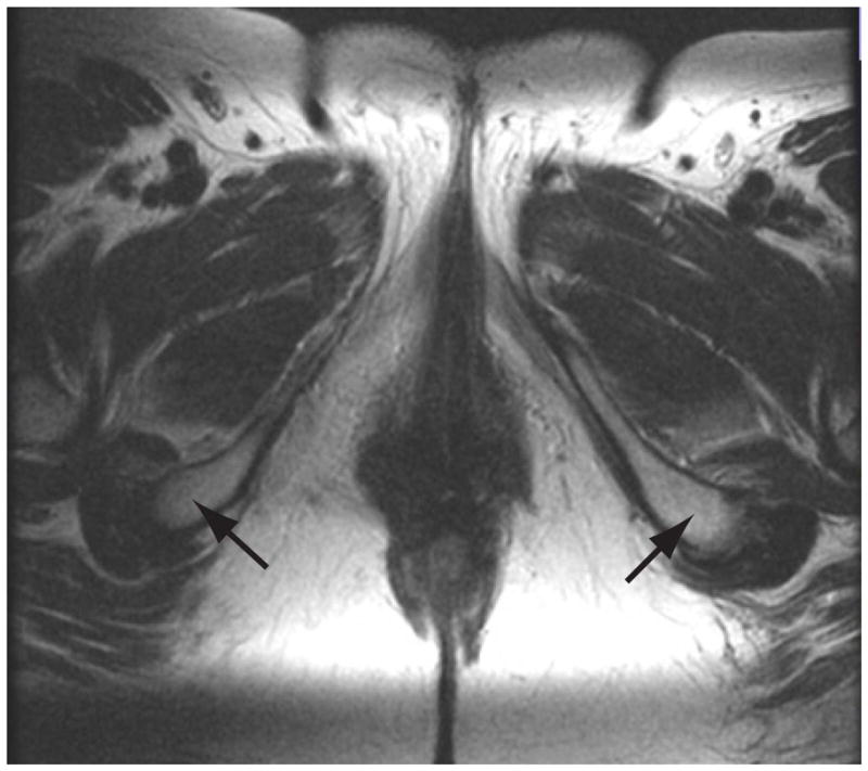
Figure 1.


MRI 2D and 3D measurement of the intertuberous distance.
Figure 1a shows the MRI 2D axial source image is shown at the level of the iscial tuberosities (Arrowheads). Figure 1b shows the 3D reconstructed image, with the black arrowheads indicating the iscial tuberosities. Color legend: White: pubic bones, yellow- urethra, pink – vagina, brown – Levator ani, rose- Obturator internus, gray – symphysis bubis and coccyx.
