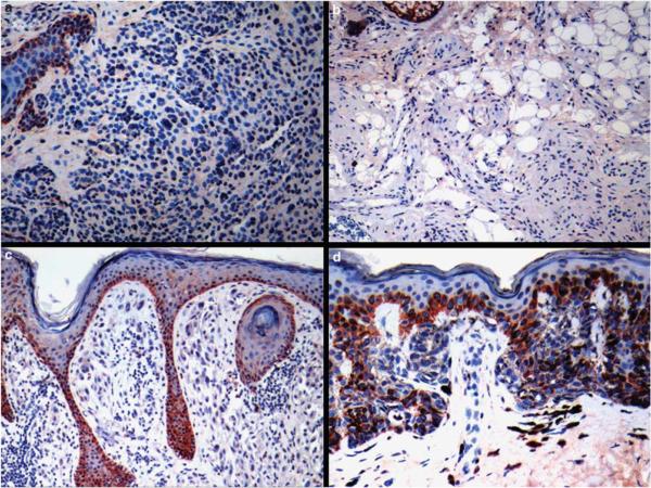
Figure 1.
S100A13 staining in melanocytic nevi: (a) absence of S100A13 immunoreactivity in a common acquired nevus. Note that basal epidermal keratinocytes are positive (upper left); (b) cytologically bland melanocytes in a neurotized dermal nevus are S100A13 negative; (c) absence of immunostaining in the spindle and epithelioid melanocytes in a common Spitz's nevus (epidermal basal keratinocytes are positive); (d) `dysplastic' nevus with mild atypia showing weak S100A13 positivity in a minority of cells.

