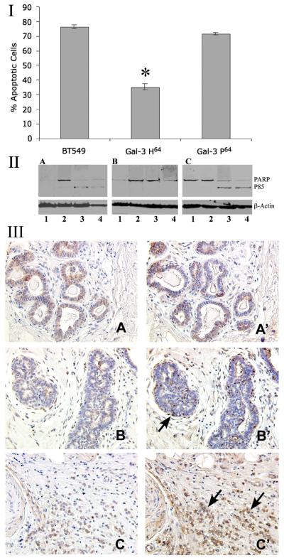
Figure 4.
Cisplatin induced apoptosis of BT-549 cells and cloned variants.
I: Percent apoptosis in cells treated with 25uM cisplatin for 72 hr at 37°C and analyzed following MTT assay. ODs in the untreated control cells were given a value of 100 and relative percent apoptosis was calculated as 100%, and the values are the average of triplicate experiments. Bars represent ±SD. * P>0.001
II: Western blot analysis of the expression of PARP in cells treated with 50 μM cisplatin after 24, 48, and 72 hr. A: BT-549; B: BT-549 Gal-3 H64; C: BT-549 Gal-3 P64. β-Actin was used as loading control. 1- Untreated; 2- 24 hr; 3- 48 hr; 4- 72 hr. Data shown is representative of three clones for each variant.
III: Immunohistochemical analysis of intact and cleaved galectin-3 in breast cancer progression. A-C: Intact protein was visualized using monoclonal anti-galectin-3 antibody TIB166; A’-C’: full length plus cleaved galectin-3 using polyclonal galectin-3 antibody hL31. For detailed description see(10). A-A’: Normal breast tissue; B-B’: Ductal hyperplasia; C-C’: Infiltrating lobular carcinoma. Arrows in B’ and C’ indicate the cleaved protein. Representative pictures are presented from a study of 5 normal breast tissues, 10 lobular hyperplasia and 8 infiltrating lobular carcinoma cases. x200

