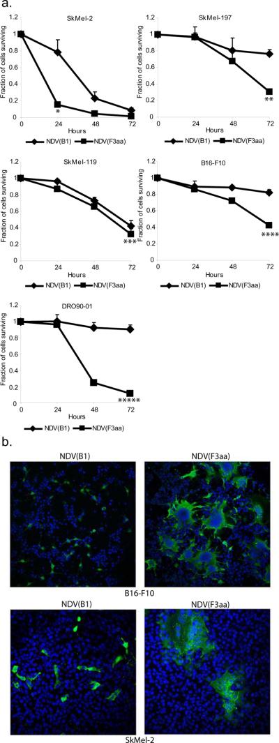
Figure 1. Recombinant NDV with enhanced fusogenic protein effectively lyses human and murine melanoma cell lines.
(a). Cell lines (5×10⁵ cells) were infected at MOI 0.1 in triplicate and LDH release assays were performed at 24, 48, and 72 hours. Percentage of cells surviving at 24, 48, and 72 hours is shown (*p=0.018, **p=0.005, ***p=0.067, ****p=0.01, *****p=0.0009). (b). Syncytia formation by the NDV(F3aa) virus. Cell lines tested in (a) were infected with NDV(B1) and NDV(F3aa) at MOI 0.01, fixed after 24 hours, and stained with dapi (blue) and anti-NDV polyclonal serum (green). Representative images from SkMel-2 and B16-F10 cells are shown.

