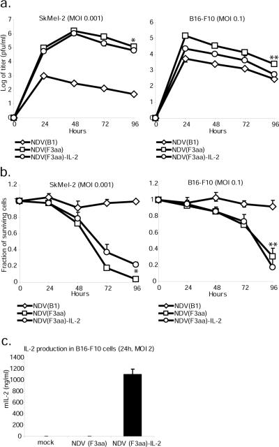
Figure 2. Recombinant NDV(F3aa)-IL-2 replicates in melanoma cells and expresses IL-2.
(a). SkMel-2 and B16-F10 cells were infected at the indicated MOI's and the viral titers in the supernatants were assessed at 24, 48, 72, and 96 hours. Statistical significance in titer difference between NDV(F3aa) and NDV(F3aa)-IL-2 was determined by Student's t-test (*p=0.009, **p=0.015). (b). B16-F10 cells (right panel) and SkMel-2 cells (left panel) were infected with NDV(B1), NDV(F3aa), and NDV(F3aa)-IL-2 viruses at the indicated MOI's. Cytotoxicity was assessed at 24, 48, and 72 hours by LDH release assays (*p=0.01, **p=0.39). Lower MOI's were used in SkMel-2 cells due to higher susceptibility of the cells to NDV. (c). B16-F10 cells were infected at MOI 2 with NDV(B1), NDV(F3aa), and NDV(F3aa)-IL-2 viruses and the supernatants were collected 24 hours post-infection. Murine IL-2 production was determined by serial dilution of supernatants and ELISA.

