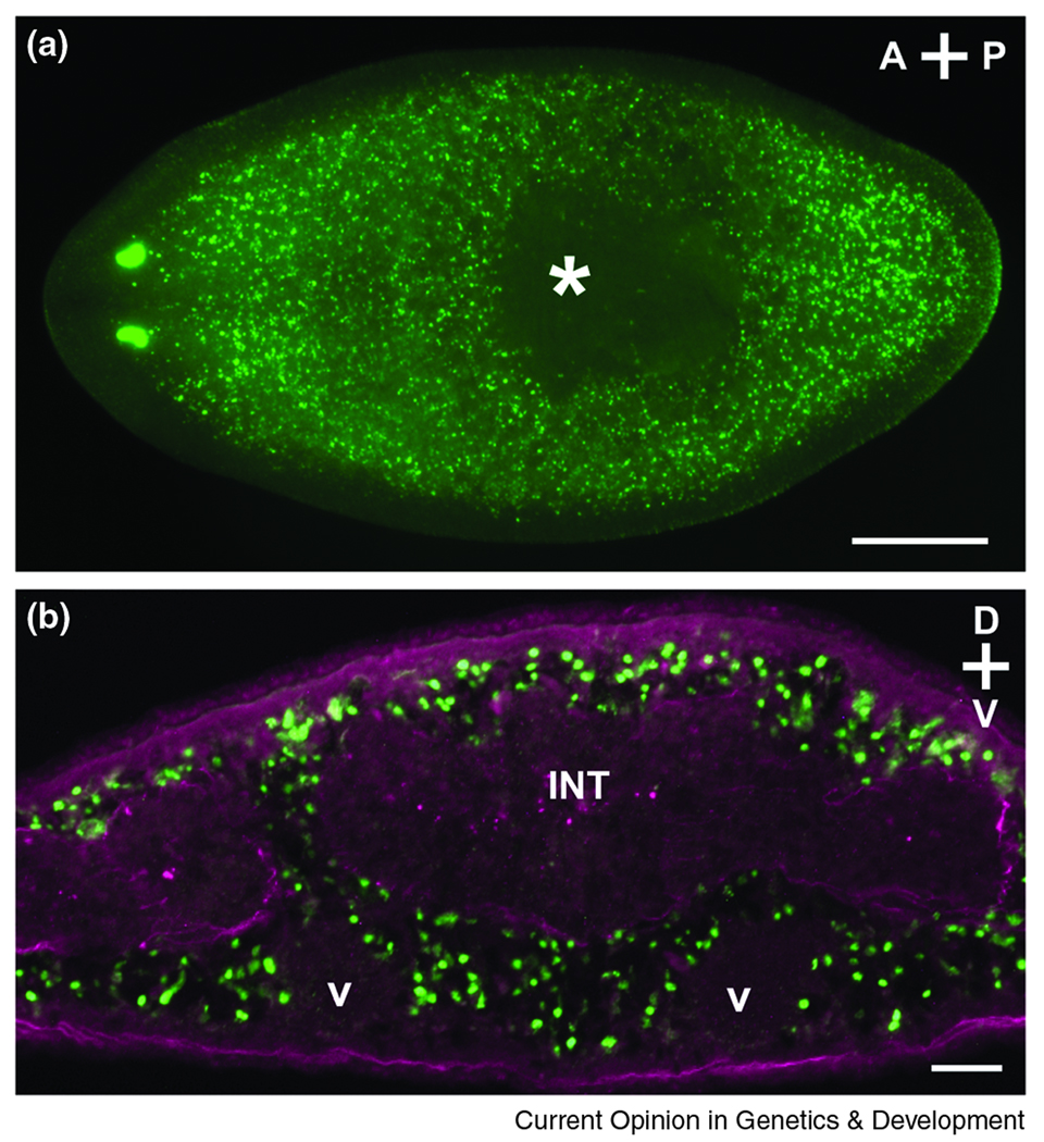
Figure 1. Planarian neoblasts.
(A) Distribution of neoblasts in an intact animal 24h after BrdU incorporation (green). S-phase cells do not reside anterior to the photoreceptors or within the pharynx (asterisk), and fragments amputated from these regions do not regenerate [1,25]. Dorsal view; anterior is to the left. Scale bar, 0.5 mm. (B) In cross section (anterior to the pharynx), neoblasts (green) are distributed mesenchymally around differentiated tissues such as the intestine (INT) and ventral nerve cords (V). Enteric and outer body wall muscles are labeled in magenta. Scale bar, 0.05 mm.

