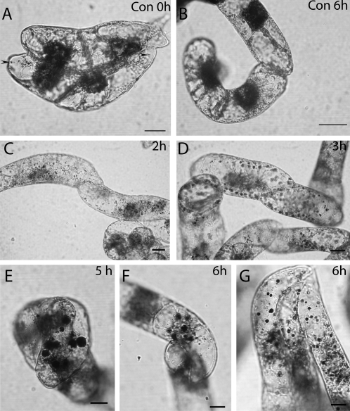
Fig. 1.
Lugol staining shows starch accumulation in BY-2 cells after BFA treatment. Scale bars=10 μm. (A and B) Control cells. Nuclei are stained brown by iodine. Only a few starch granules are visible (A, arrows). Control cells after 6 h show no starch signal at all (B). (C) Two hours after adding BFA the number of iodine-positive starch granules increases. (D) Three hours after adding the inhibitor the black starch granules are easy to observe. (E) Four hours after adding BFA in younger cells large starch granules are visible. (F and G) The size of starch granules in BY-2 cells after adding BFA increases continuously up to 6 h of treatment.

