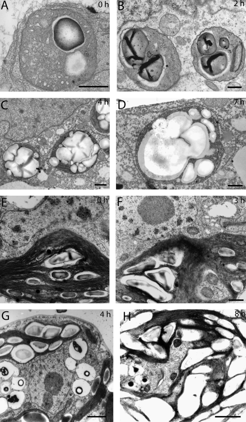
Fig. 2.
Electron micrographs showing starch accumulation in BY-2 cells (A–F, scale bars=500 nm) and Chlamydomonas noctigama plastids (E–G, scale bars=1000 nm) treated with BFA (10 μg ml−1). (A) Untreated BY-2 cells show typical patterns of proplastids. Undefined membranes are present in the stroma. Plastids can accumulate starch in the matrix typical of amyloplasts. (B) After 2 h BFA treatment nearly all plastids contain starch granules. (C) At 4 h after BFA treatment starch grains occupy most of the plastid volume. (D) After 9 h, fusion of the starch grains is more or less complete and the plastid matrix is completely filled with starch. (E) A typical Chlamydomonas chloroplast with a number of small starch granules located in the interthylakoid spaces. (F) At 3 h after adding BFA, starch seems to accumulate. Starch granules are considerably larger compared with the control. (G) A 4 h BFA treatment leads to a significant increase of starch within the plastid. (H) After 8 h the interthylakoidal spaces are filled with a number of large starch grains.

