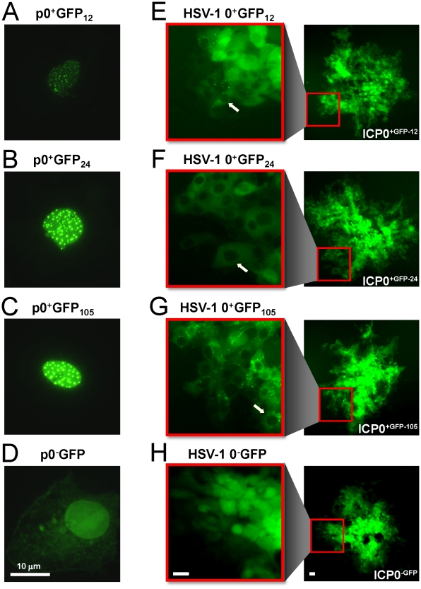
Figure 2. GFP-tagged ICP0 is predominantly cytoplasmic in HSV-1 infected cells.
(A) ICP0+GFP-12, (B) ICP0+GFP-24, (C) ICP0+GFP-105, or (D) ICP0−GFP as seen in Vero cells 12 hours post transfection with the plasmids p0+GFP12, p0+GFP24, p0+GFP105, or p0−GFP, respectively. Each plasmid was co-transfected with the plasmid pBHad.CMV-VP16 to induce the ICP0 promoter and expression of GFP-tagged ICP0. (E) ICP0+GFP-12, (F) ICP0+GFP-24, (G) ICP0+GFP-105, or (H) ICP0−GFP as seen in Vero cells 40 hours post inoculation with the HSV-1 recombinant viruses 0+GFP12, 0+GFP24, 0+GFP105, or 0−GFP, respectively. In each panel, an entire plaque is shown on the right, and one edge of the plaque is magnified on the left. White arrows denote HSV-1 infected cells in which GFP-tagged ICP0 was abundant in the cytoplasm, but was not evident in the nuclei of HSV-1 infected cells. The scale bar denotes a distance of 10 µm.

