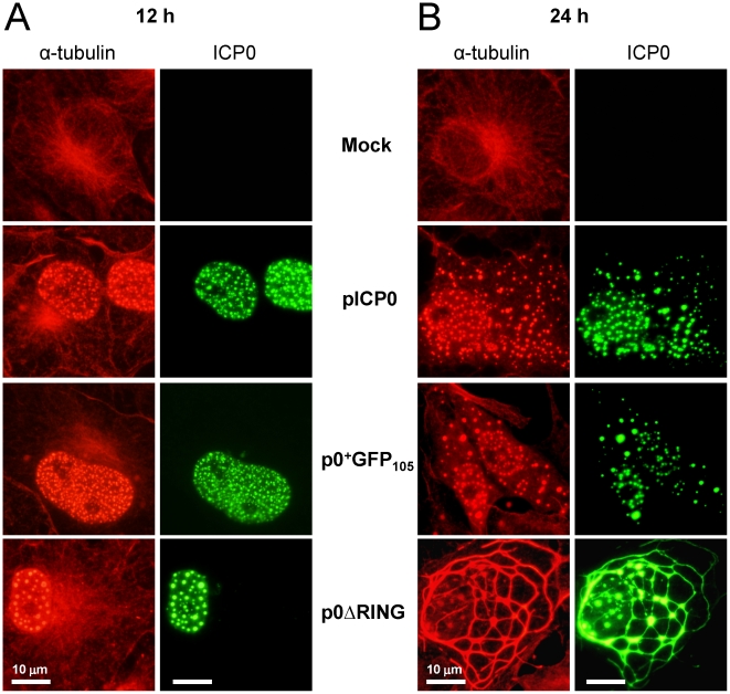
Figure 9. ICP0 is sufficient to trigger dispersal of host cell microtubule networks.
Vero cells were mock-transfected or were transfected with a VP16-expressing plasmid and pICP0, p0+GFP105, or p0ΔRING. At (A) 12 hours post-transfection or (B) 24 hours post-transfection, mock- and pICP0-transfected cells were fixed and stained with antibodies against α-tubulin (rabbit IgG) and ICP0 (mAb H1083). Cells transfected with p0+GFP105 or p0ΔRING were fixed and stained to visualize α-tubulin, and the ICP0ΔRING and ICP0+GFP-105 proteins were visualized using their GFP fluorophores. The scale bar denotes a distance of 10 µm.

