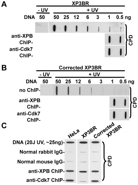
Figure 4. Enrichment of UV-induced photolesions by anti-XPB and anti-Cdk7 ChIP in XP3BR and XPG cDNA-corrected XP3BR cells.
The unirradiated or UV-irradiated (20 J/m2) fibroblasts were cultured for 1 h to allow DNA repair before fixation. The soluble chromatin preparation, ChIP and DNA isolation were carried out as described in Figure 1C. (A) and (B) Predetermined amount (0.5 and 1.0 ng) of ChIP-recovered DNA was used for Immunoslot-blot analysis of CPD in anti-XPB and anti-Cdk7 ChIP-recovered DNA from XP3BR (A) or XPG cDNA-corrected XP3BR (B) cells. (C) The ChIP-recovered DNA from the same amount of soluble chromatin (500 µg in protein) was used for Immunoslot-blot analysis of CPD in HeLa, XP3BR and XPG cDNA-corrected XP3BR cells. Genomic DNA samples isolated from UV-irradiated cells were used as positive controls.

