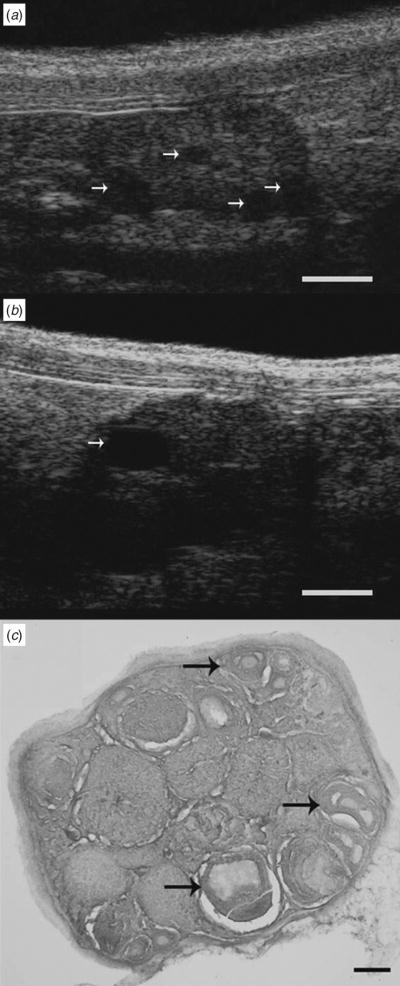
Fig. 1.
Ultrasound biomicroscopy and histological sections of a mouse ovary depicting follicles at different stages of development. Arrows on UBM images indicate ovarian follicles ranging from (a) 290 to 440 μm in diameter and (b) a preovulatory-sized follicle measuring 730 μm in diameter. (c) The arrows in the histological image indicate ovarian follicles ranging from 220 to 385 μm in diameter. Scale bar: (a, b) 1000 μm, (c) 200 μm.

