
Fig. 1.
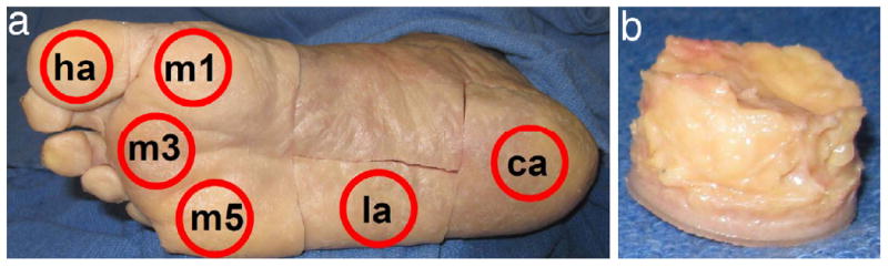
Specimen locations (a) at the hallux (ha), first, third, and fifth metatarsal heads (m1, m3, and m5), lateral midfoot (la), and calcaneus (ca) as well as (b) a typical plantar tissue specimen before skin removal.
Official websites use .gov
A
.gov website belongs to an official
government organization in the United States.
Secure .gov websites use HTTPS
A lock (
) or https:// means you've safely
connected to the .gov website. Share sensitive
information only on official, secure websites.
Specimen locations (a) at the hallux (ha), first, third, and fifth metatarsal heads (m1, m3, and m5), lateral midfoot (la), and calcaneus (ca) as well as (b) a typical plantar tissue specimen before skin removal.