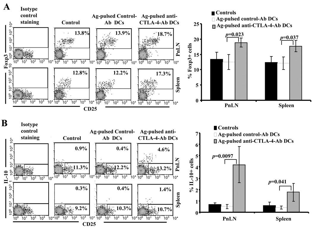
FIGURE 6.
Treatment of NOD mice with Ag-pulsed anti-CTLA-4-Ab DCs results in the induction of Foxp3+ and IL-10+ T cells. Eight week old euglycemic female NOD mice were left untreated or treated with antigen-pulsed control or anti-CTLA-4 Ab coated DCs as described above for Fig. 3. On day 15 post-treatment, treated and untreated control mice were euthanized and freshly isolated spleen and pancreatic LN cells were stained for surface and intra-cellular markers using fluorochrome labeled Abs and analyzed by FACS. CD4+ population was gated for both panels. Representative scatter plots and percentage values for A) CD4+Foxp3+ and B) CD4+IL-10+ T cells (left panels) and mean±SD of the percentage values obtained using cells from at least 6 mice/group (right panels) are shown.

