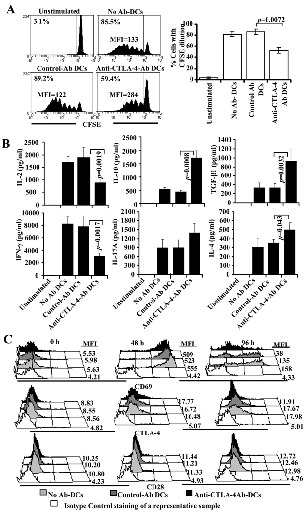
FIGURE 8.
DC directed enhanced engagement of CTLA-4 does not down-regulate CD28 expression, but promotes regulatory cytokine production leading to suppression of activated T cells. A) Non-pulsed or BDC peptide-pulsed DCs without (none), or with control or anti-CTLA-4 Ab coating were incubated with CFSE labeled purified CD4+ T cells from NOD.BDC2.5 TCR-Tg mice. Cells from these cultures were tested for CFSE dilution by FACS on day 4 after staining with PE-labeled anti-CD4 Ab. CD4+ T cells were gated for this panel. Representative histogram plots and percentage values of CD4+ T cells with CFSE dilution (left panel) and mean±SD of values from two independent assays carried out in triplicate (right panels) are shown. B) Supernatants collected from 72 h parallel cultures were tested for cytokines by ELISA. Mean±SD of values from three separate experiments carried out in triplicate are shown for panel B. C) Purified BDC2.5 T cells were cultured with BDC peptide-pulsed non-coated (control DCs) or Ab coated DCs for different durations, stained with fluorochrome labeled CD4, CD28, CTLA-4, CD69 specific Abs, and analyzed by FACS. The CD4+ population was gated for this panel. Each sub-panel shows a representative sample stained using an isotype control Ab and overlay of samples stained using Ab for a specified marker, and mean fluorescence intensity (MFI) value for each sample. The assay was repeated twice in triplicate with similar results.

