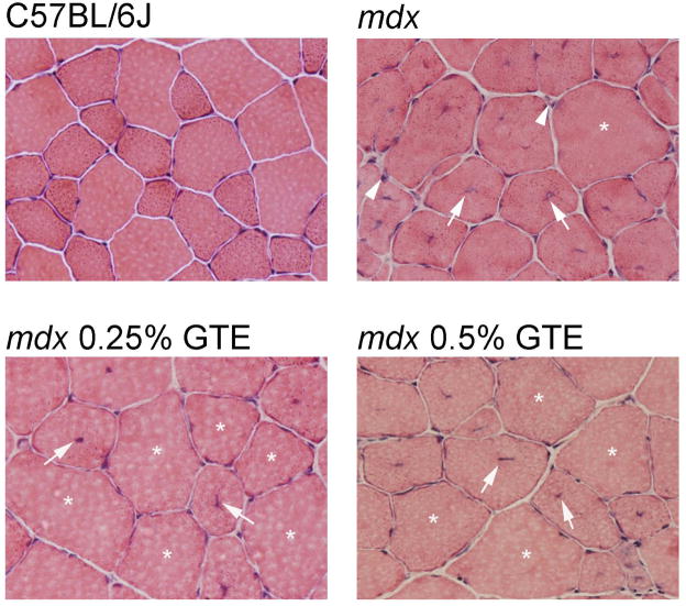
Figure 4.
Histopathology of TA muscles for C57BL/6J, mdx and GTE treated mdx mice age 42 days. C57BL/6J muscle sections depicting normal fiber morphology for this age. Untreated mdx sections showing a morphologically normal fiber (asterisks), regenerating fibers (arrow), and immune cell infiltration (arrowhead). Treated mdx sections from 0.25% GTE and 0.5% GTE had altered muscle pathology with an increase in morphologically normal fibers (asterisks) and fewer regenerating fibers (arrow). 400X H&E.

