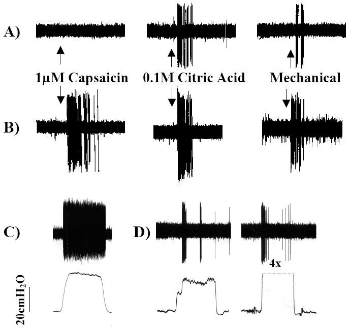
Figure 1.

Representative extracellular recordings from the vagal afferent nerve subtypes innervating the airways and lungs. A) The cough receptors innervate the larynx, trachea and mainstem bronchi. They are insensitive to capsaicin, airway smooth muscle contraction (not shown) and distending or collapsing airway luminal pressures (not shown) but are activated by punctate mechanical stimulation and acid. When activated, these afferent nerves initiate coughing. B) Bronchopulmonary C-fibers terminate throughout the airways and lungs. C-fibers are less sensitive to mechanical stimulation than other airway afferent nerves but are activated by chemical stimuli such as capsaicin, bradykinin, acid and adenosine. When activated, C-fibers initiate coughing, changes in respiratory pattern and autonomic reflexes (e.g. airway smooth muscle contraction, mucus secretion. C-fiber subtypes have been described. C) Slowly adapting receptors are active during the dynamic and static phases of lung inflation. Slowly adapting receptor activation initiates respiratory slowing but does not initiate coughing. D) Rapidly adapting receptors are active during the dynamic phases of lung inflation and deflation and are activated by airway smooth muscle contraction/ bronchospasm and lung collapse/ negative airway luminal pressures. Rapidly adapting receptor activation initiates parasympathetic reflexes such as mucus secretion and airway smooth muscle contraction as well as tachypnea.
Figures are reproduced with permission from Canning et al., 2004 (49). Canning BJ, Mazzone SB, Meeker SN et al. Identification of the tracheal and laryngeal afferent neurones mediating cough in anaesthetized guinea-pigs. J Physiol 2004;557:543-58.
