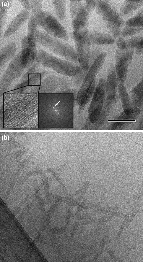
Fig. 2.
Isolated chlorosomes embedded in an amorphous ice layer give hints of the overall and internal structure. a Overview of unstained chlorosomes of Chlorobium tepidum. The inset shows a fine parallel spacing of lamellae, its calculated diffraction pattern indicates a strong diffraction spot equivalent with a 2.1-nm lamellar spacing. b Unstained ice-embedded chlorosomes of Chloroflexus aurantiacus (phylum Chloroflexi or filamentous anoxygenic phototrophs). The ice layer has been prepared over a holey-carbon film, which is visible at the lower left side. Size bar for both frames equals 100 nm

