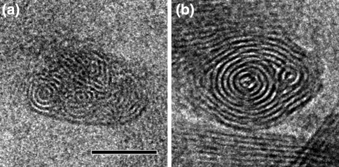
Fig. 3.
End-on views of chlorosomes of Chlorobaculum tepidum, fixed in a vertical position in an amorphous ice layer. Cryo-EM reveals the packing of the lamellae. a Packing in the wild-type with some of the lamellae in concentric rings, others in a more irregular association. b Packing in the bchQRU mutant, showing a more regular multi-cylindrical organization. See also (Oostergetel et al. 2007) for further images. Size bar equals 25 nm

