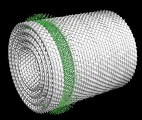
Fig. 7.
Cylindrical model of the packing of concentric lamellae in the Chlorobaculum tepidum bchQRU mutant, based on distances as observed by electron microscopy and solid-state NMR spectroscopy (Ganapathy et al. 2009). The spacing between layers is 2.1 nm. The green band indicates the position of individual Bchl molecules in four stacks of syn-anti dimers. In the wild-type chlorosomes, the stacks run in the direction of the cylinder axis

