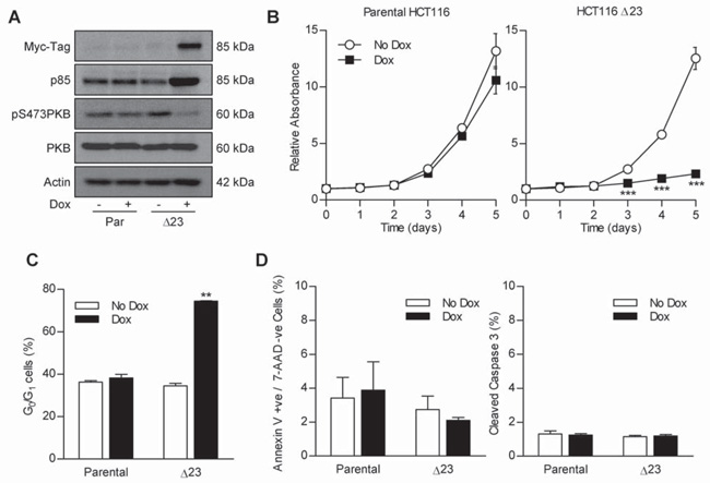
Figure 3. MycΔp85α expression inhibits PI3K signaling and causes a cell-cycle arrest in HCT116 cells.
A Parental HCT116 and clone Δ23 cells were grown in the presence or absence of 0.5 mg.ml−1 Dox for 24 h and lysed. Lysates were assayed for the level of Myc-tagged protein, p85, phospho-PKB and total-PKB by western blotting. B Cells were seeded into 96 well plates and after 24 h treated with 0.5 mg.ml−1 dox or left untreated. A plate was harvested every 24 h for five days and the amount of protein in each well relative to day 0 was determined by SRB staining. *** p<0.001 according to two-tailed unpaired t-test compared to corresponding no dox treatment. C Cells were grown in the absence or presence of dox for 24 h and harvested by trypsinisation and fixed in 70% ethanol. The cell-cycle profile was determined and % of cells in the G0/G1 stage of the cell-cycle calculated as in the previous figure. ** p<0.01 according to two-tailed unpaired t-test compared to all other groups. D Cells were grown in the absence or presence of dox for 24 h. The % annexin V +ve / 7-AAD −ve cells and the % cleaved caspase 3 was determined as in the previous figure. All graphs represent the mean from three independent experiments +/− S.E.M. Blots are representative examples from three independent experiments.

