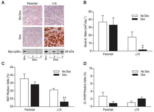
Figure 5. MycΔp85α expression inhibits xenografts tumor growth rate and Ki67 staining but has no effect on the level of PARP cleavage.
Parental HT29 cells and clone Δ15 cells were grown as tumor xenografts on Balb/c-nude mice and tumors were measured three times a week by callipers. Once tumors reached 300 mm3 mice were switched onto feed containing dox or control-feed. Four animals were sacrificed three days after being switched onto dox/control-feed and their tumors were bisected and either snap frozen or fixed in 10% formalin. A further six mice from each group were used for tumor growth analysis. A Top Panel – Tumors were assayed for the presence of Myc-tagged proteins by IHC: Bottom Panel – Tumor lysates were assayed for the presence of MycΔp85α by western blotting. +ve = positive control of a dox induced clone Δ15 cell lysate B The mean (+/− S.E.M) of the tumor growth rate from each group of mice after the tumor reached 300 mm3. C and D Tumors were stained for Ki67 (C) and cleaved PARP (D) by IHC and the percentage of positive cells counted. Data represents the mean +/− S.E.M. * p<0.05, ** p<0.01 compared to all other groups according to two-tailed unpaired t-test.

