
Figure 6. MycΔp85α expression does not enhance oxaliplatin-induced or etoposide-induced apoptosis.
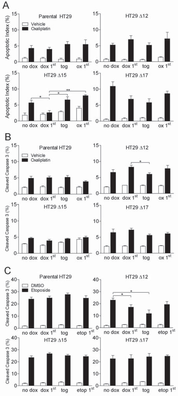
A and B Cells were treated for 48 h with a concentration of oxaliplatin which gave 5-10% apoptosis or vehicle control (PBS). This was either the only treatment the cells received (no dox) or the cells were also treated with dox 24 h prior to oxaliplatin (dox 1st), at the same time as oxaliplatin (tog) or 24 h after oxaliplatin (ox 1st). 48 h after the start of oxaliplatin treatment cells were harvested by trypsinisation and either fixed in 70% ethanol or lysed. A Fixed cells were stained with DAPI and the percentage of cells with an apoptotic nuclei were counted. B The percentage of cleaved-caspase 3 within lysates was determined as described in previous figures. C Cells were treated for 48 hours with 100 μM etoposide or DMSO equivalent. This was either the only treatment the cells received (no dox) or the cells were also treated with dox 24 h prior to etoposide (dox 1st), at the same time as etoposide (tog) or 24 h after etoposide (etop 1st). 48 h after the start of etoposide treatment cells were harvested by trypsinisation and lysed. The percentage of cleaved-caspase 3 within lysates was determined using Mesoscale Discovery cleaved- and total-caspase 3 duplex plates. All graphs represent the mean from three independent experiments +/− S.E.M. * p<0.05, ** p<0.01 according to two-tailed unpaired t-test.