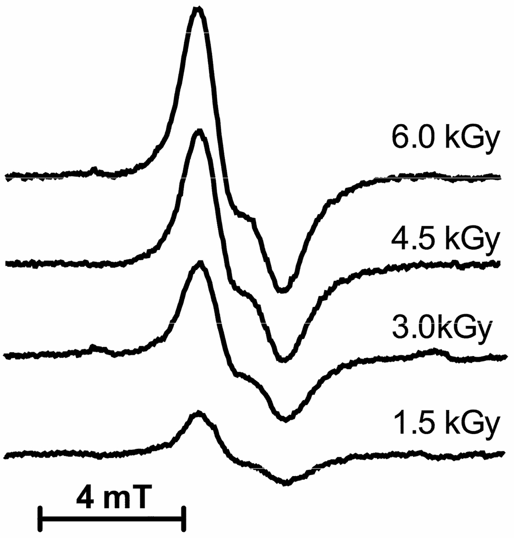
Figure 4.
Representative EPR spectra are shown for Mlut films hydrated to Γ = 2.5. First derivative spectra were recorded at 4 K using Q-band frequency and microwave power attenuation set at 50 db. The dose is shown to the right of each spectrum. The concentration was determined by comparison with an EPR signal from ruby (not shown) that serves as an internal standard. The radical concentrations were plotted as a function of dose in Figure 5.

